The Ultimate Guide To Rhinoplasty Surgery Austin
Table of ContentsAustin Rhinoplasty Can Be Fun For EveryoneRhinoplasty Surgery Austin for DummiesRhinoplasty Surgery Austin Can Be Fun For Anyone10 Simple Techniques For Rhinoplasty Surgery Austin
Ultrasonic nose surgery uses piezoelectric instruments (scrapers rasps, saws) that affect just the bones and the stiff cartilages through ultrasonic vibrations, as the instruments utilized in oral surgery. Using piezoelectric instruments requires a more prolonged method than the isial one, allowing to imagine the whole bony vault, to improve it with rhinosculpture or to activate and support bones after controlled osteotomies.Normally, the plastic surgeon first separates the nasal skin and the soft tissues from the osseo-cartilagenous nasal framework, and then improves them, sutures the cuts, and uses either an external or an internal stent, and tape, to paralyze the freshly rebuilded nose, and so facilitate the healing of the surgical cuts.
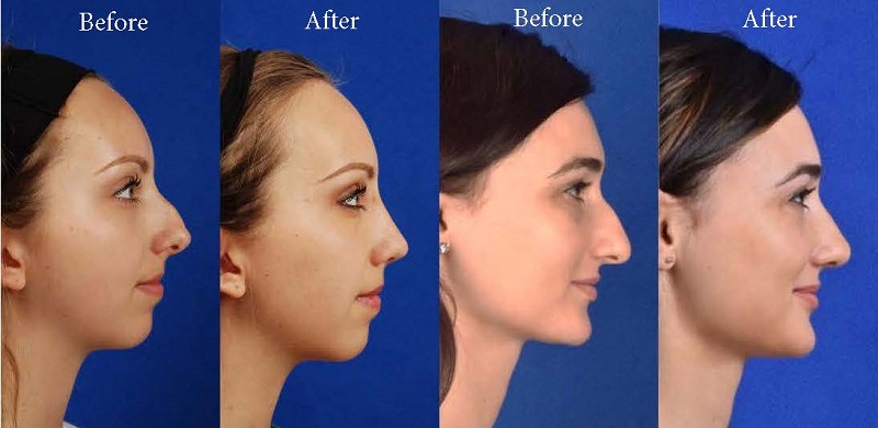
To record the "before-and-after" physiognomies of the nose and the face of the patient, the particular visual point of views needed are pictures of the nose seen from the anteroposterior (front-to-back) perspective; the lateral view (profiles), the worm's- eye view (from listed below), the bird's- eye view (overhead), and three-quarter-profile views. Photo A. Open rhinoplasty: At rhinoplasty's end, after the cosmetic surgeon has actually sutured (closed) the incisions, the fixed (brand-new) nose will be dressed, taped, and splinted immobile to allow the uninterrupted healing of the surgical incisions.
Photograph B. Open rhinoplasty: The brand-new nose is prepared with paper tape in order to receive the metal nasal-splint that will immobilize it to maintain its appropriate shape as a brand-new nose. Photo A. Open nose job: Pre-operative, the standards (purple) made sure the cosmetic surgeon's precise cuts in cutting the nasal defect correction strategy.
Get This Report on Rhinoplasty Austin

Open nose surgery: After the initial taping of the nose, a custom-made, metal nasal-splint, created, cut, and formed by the cosmetic surgeon, is emplaced to immobilize and safeguard the tender tissues of the new nose during convalescence. Picture D. Open nose surgery: The taping, emplacement of the metal splint, and dressing of the brand-new nose finish the rhinoplasty procedure - austin rhinoplasty.
Photo 2. Open nose surgery: The right lower lateral cartilage (blue) is exposed for correction. Photo 1. Open rhinoplasty: The columellar cut marked as a red-dot guideline, will assist the surgeon in the accurate suturing of the nose. Photo 4. Rhinoplastic correction: A nasal-hump excision strategy; the black line marks the dorsal aircraft of the brand-new nose.
Open nose surgery: the nasal pointer is sutured to narrow the nose. Photograph 1. Open nose job: The incisions are endonasal (in the nose), and hence are hidden. The skin-incision to the columella help the cosmetic surgeon in exactly suturing to conceal the scarexcept for the columellar cut (red-dot guideline) across the nasal base.
Picture 2. Open rhinoplasty: The nasal interior. The scissors suggest the lower lateral cartilage (blue), which is among the wing-shaped cartilages that adhere the pointer of the austin rhinoplasty nose. The rugged red delineation indicates the place of the columellar incision. When the skin has actually been lifted from the bone-and-cartilage structure, the cosmetic surgeon performs the nasal correction tasks.
The 5-Second Trick For Rhinoplasty Surgery Austin
Open nose job: To narrow the pointer of a too-wide nose, the surgeon first figures out the reason for the excess nasal width. The stitch being emplaced will narrow the pointer of the nose. The red delineation suggests the edge of the nose-tip cartilage, which is narrowed when the cosmetic surgeon tightens the folded cartilage apex.
Picture 4. Nasal bulge excision: The black delineation shows the preferred nose-reduction outcome: a straight nose. The nasal bulge is bone (red) above the scalloped grey line, and cartilage (blue) below the scalloped grey line. The cosmetic surgeon cuts the cartilage part of the bulge with a scalpel, and chisels the bone portion with an osteotome (bone chisel).
Although many modification nose surgery procedures are "open approach", such a correction is more technically made complex, typically because the nasal assistance structures either were warped or destroyed in the main nose job; thus the cosmetic surgeon should re-create the nasal support with cartilage grafts harvested either from the ear (auricular cartilage graft) or from the rib cage (costal cartilage graft).
In reconstructive nose surgery, the defects and defects that the plastic surgeon encounters, and should restore to typical function, kind, and appearance consist of broken and displaced nasal bones; interfered with and displaced nasal cartilages; a collapsed bridge of the nose; congenital flaw, injury (blunt, penetrating, blast), autoimmune condition, cancer, intranasal drug-abuse damages, and stopped working main nose job outcomes.
Not known Details About Rhinoplasty Austin
When cartilage is interrupted, suturing for re-suspension (structural assistance), or using cartilage grafts to camouflage an anxiety enable the re-establishment of the normal nasal shape of the nose for the client. When the bridge of the nose is collapsed, rib-cartilage, ear-cartilage, or cranial-bone grafts can be used to restore its structural stability, and thus the visual connection of the nose.